Label the specific serous membranes and cavity of the heart. – Labeling the specific serous membranes and cavity of the heart is an essential aspect of understanding the anatomy and physiology of the heart. This article provides a comprehensive overview of these structures, their functions, and their clinical significance.
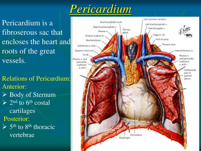
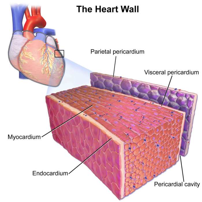
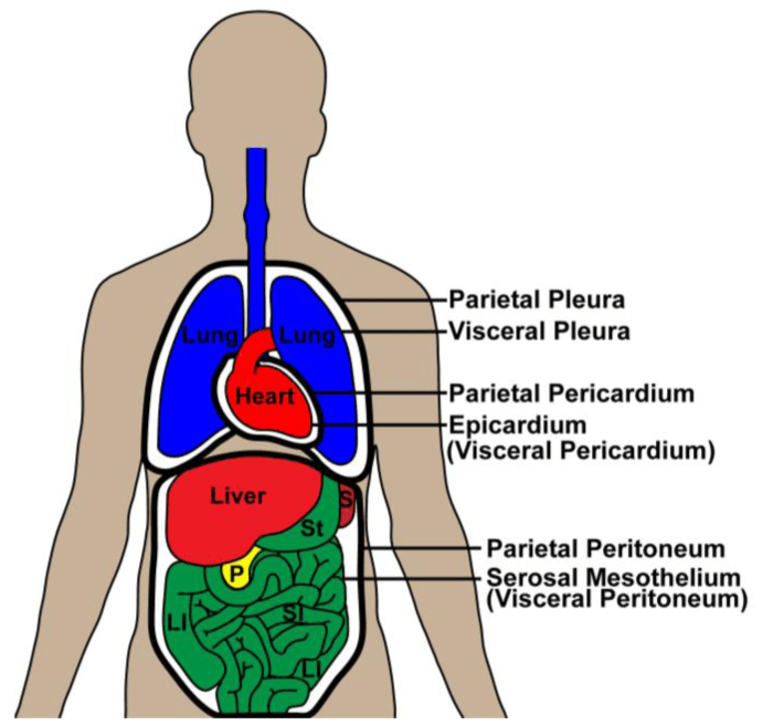
The heart is enclosed within a double-layered serous membrane known as the pericardium. The fibrous pericardium forms the tough outer layer, while the serous pericardium consists of two layers: the parietal layer lines the fibrous pericardium, and the visceral layer, also known as the epicardium, covers the surface of the heart.
Serous Membranes and Cavities of the Heart: Label The Specific Serous Membranes And Cavity Of The Heart.

The heart is enclosed within a double-layered serous membrane called the pericardium. The serous membranes are composed of two layers: a parietal layer that lines the cavity and a visceral layer that covers the organ. Between the two layers is a serous cavity filled with a thin film of serous fluid that reduces friction during heart contractions.
Pericardium
The pericardium consists of two layers: a tough outer fibrous pericardium and a thin inner serous pericardium. The fibrous pericardium protects the heart from external forces and provides attachment points for the great vessels. The serous pericardium consists of a parietal layer that lines the pericardial cavity and a visceral layer, called the epicardium, that covers the heart.
The pericardial cavity is a potential space between the parietal and visceral layers of the serous pericardium. It contains a small amount of serous fluid that lubricates the heart and reduces friction. The pericardial cavity is clinically significant because it can become inflamed, a condition known as pericarditis.
Epicardium, Label the specific serous membranes and cavity of the heart.
The epicardium is the visceral layer of the serous pericardium. It is a thin, transparent membrane that covers the surface of the heart. The epicardium is continuous with the endocardium, the inner lining of the heart chambers.
The epicardium contains a network of blood vessels that supply the heart muscle with oxygen and nutrients. Inflammation of the epicardium, known as epicarditis, can lead to chest pain and other symptoms.
Myocardium
The myocardium is the middle layer of the heart wall. It is composed of cardiac muscle fibers, which are arranged in a complex network that allows the heart to contract and pump blood.
The myocardium is supplied with blood by the coronary arteries. The coronary arteries branch off the aorta and supply oxygen and nutrients to the heart muscle. Blockage of the coronary arteries can lead to a heart attack.
Endocardium
The endocardium is the inner lining of the heart chambers. It is a thin, smooth membrane that prevents blood from leaking out of the heart.
The endocardium is composed of endothelial cells, which are specialized cells that line the blood vessels. Endothelial cells help to regulate blood flow and prevent the formation of blood clots.
Damage to the endocardium, such as in endocarditis, can lead to heart failure and other serious complications.
FAQs
What is the function of the pericardial cavity?
The pericardial cavity contains a small amount of fluid that lubricates the heart and reduces friction during its contractions.
How is the epicardium continuous with the endocardium?
The epicardium and endocardium are continuous at the atrioventricular valves and the openings of the great vessels.
What is the clinical significance of endocardial damage?
Endocardial damage can lead to endocarditis, a serious infection of the inner lining of the heart chambers.

